A new operative technology has been introduced to thyroid surgery at Cleveland Clinic London: the Zeiss operative microscope, providing surgeons with magnified, brilliantly illuminated visualisation of the recurrent laryngeal nerve and parathyroid glands during thyroid dissection — with the aim of reducing complications and increasing the delicacy and accuracy of the operation.
The two most significant complications of total thyroidectomy — injury to the recurrent laryngeal nerve and damage to the parathyroid glands — both result from a failure to identify and protect structures that are often small, variably positioned, and easily confused with surrounding tissue. The recurrent laryngeal nerve may be as fine as 1–2mm in calibre. The parathyroid glands, each weighing approximately 40mg, can be mistaken for fat or lymphatic tissue. In conventional thyroid surgery performed with loupe magnification — or no magnification at all — these structures are dissected under visual conditions that are inherently limited.
The Zeiss operative microscope addresses this limitation directly. Offering steplessly adjustable magnification typically ranging from 4x to 16x, with coaxial xenon or LED illumination and an optically superior depth of field, the microscope transforms the operative field into a landscape in which individual nerve fascicles, vascular pedicles, and parathyroid tissue planes are resolved with a clarity that is simply not achievable with loupe magnification alone.
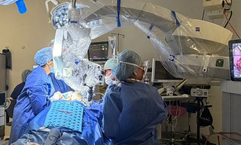
"The Zeiss microscope transforms what is visible to the surgeon during the most critical moments of thyroid dissection — the identification of the recurrent laryngeal nerve and the parathyroid glands. Structures that might otherwise be at risk become clearly defined and unambiguous."
Why magnification matters in thyroid surgery
The recurrent laryngeal nerve runs a variable course in the neck and enters the larynx in close proximity to the posterior aspect of the thyroid lobe. Its diameter may be as small as 1–2mm. A branch pattern — in which the nerve divides into two or more branches before entering the larynx — occurs in approximately 30–40% of patients and represents a particular identification challenge. Even brief inadvertent stretch, thermal proximity or contact from a dissecting instrument can cause transient or permanent vocal cord palsy.
The parathyroid glands present a parallel challenge. Their average dimensions are approximately 6mm × 4mm × 2mm, and their colour — yellow-brown, closely resembling fat or lymph node tissue — makes identification under poor lighting or low magnification genuinely difficult. Inadvertent excision of a parathyroid gland, or interruption of its delicate blood supply during dissection, is the primary mechanism of postoperative hypoparathyroidism.
Conventional loupe magnification used in thyroid surgery typically provides 2.5× to 4.5× magnification — useful, but limited. The Zeiss operative microscope offers stepless magnification from approximately 4× to 16×, with vastly superior depth of field, contrast resolution, and coaxial illumination that eliminates the shadows inherent to loupe systems. At higher magnifications, individual nerve fascicles are visible, fat planes adjacent to parathyroid glands resolve clearly, and the delicate vascular pedicles supplying the parathyroids can be identified and preserved with a precision not achievable by any other means.
Combined with continuous RLN monitoring and medial-to-lateral dissection
The Zeiss microscope joins two other recent additions to the operative approach to total thyroidectomy at Cleveland Clinic London: continuous intraoperative recurrent laryngeal nerve monitoring (C-IONM) and the medial-to-lateral dissection technique described in a recent blog post. Together, these three elements form a layered approach to risk reduction in thyroid surgery.
Continuous RLN monitoring provides real-time electrophysiological feedback about nerve function throughout the dissection. The medial-to-lateral technique protects the parathyroid vascular territory by approaching it in the correct anatomical sequence. The Zeiss microscope adds the optical precision to execute both with a level of visual clarity that conventional magnification cannot match. These technologies are complementary rather than redundant — each addressing a different mechanism by which complications arise.
The patient benefit
For patients undergoing total thyroidectomy at Cleveland Clinic London, the introduction of operative microscopy represents a meaningful advance in the safety and precision of their surgery. The two most feared consequences of thyroid surgery — a permanently changed voice from nerve injury, and lifelong calcium-dependence from parathyroid damage — both become less likely when the structures responsible are identified and dissected under conditions of maximum visual clarity.
This is not a technique that changes what is done in thyroid surgery. It changes what can be seen — and therefore what can be protected — with greater confidence at every step of the operation.